
' 
SCIENTIFIC SCORE
Most Likely Effective
Based on 31 Researches
7.7
USERS' SCORE
Very Good
Based on 1 Reviews
8.5
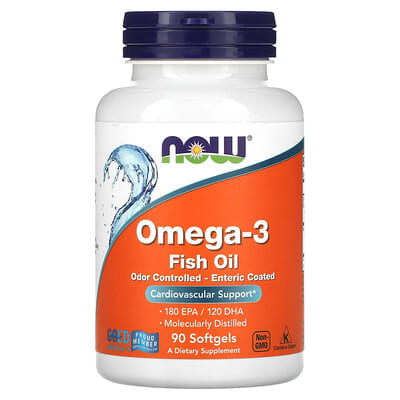
Supplement Facts
Serving Size: 1 Softgel
Amount Per Serving
%DV
Calories
10
Total Fat
1 g
1%**
Saturated Fat
< 0.5 g
2%**
Polyunsaturated Fat
0.5 g
†
Fish Oil Concentrate
1 g (1,000 mg)
†
Omega-3 Fatty Acids:
Eicosapentaenoic Acid (EPA)
180 mg
†
Docosahexaenoic Acid (DHA)
120 mg
†
Top Medical Research Studies
9
DHA-PS promotes liver recovery
DHA-enriched phosphatidylserine ameliorates cyclophosphamide-induced liver injury via regulating the gut-liver axis.
Highly relevant to liver health
We investigated the potential of DHA-enriched phosphatidylserine (DHA-PS) in addressing liver injuries caused by cyclophosphamide in mice. By administering cyclophosphamide over five days, we created a model to simulate this liver damage. Following this, we treated the mice with different doses of DHA-PS for a week to see if it could help heal their livers.
Our findings were quite promising. The mice receiving DHA-PS experienced significant improvements in key liver health indicators, including reduced inflammation and oxidative stress. Through advanced analysis, we discovered that DHA-PS helped correct metabolic imbalances caused by cyclophosphamide, which is a crucial aspect of liver recovery.
Additionally, we observed that DHA-PS has a positive impact on the gut-liver axis. This treatment restored the intestinal barrier, reduced harmful compounds in the bloodstream, and improved overall gut microbiota health. By balancing the gut bacteria, we enhanced the overall health of the mice.
Overall, the results suggest that DHA-PS could be a valuable therapeutic option or functional food for combating liver injuries related to cyclophosphamide. This study highlights the potential benefits of DHA on liver health and underscores the importance of the gut-liver connection.
Read More
9
DHA's Role in Liver Health
Biosynthetic deficiency of docosahexaenoic acid causes nonalcoholic fatty liver disease and ferroptosis-mediated hepatocyte injury.
Study highlights DHA's importance
We explored the impact of docosahexaenoic acid (DHA), a type of omega-3 fatty acid, on the health of our liver, especially regarding nonalcoholic fatty liver disease (NAFLD). Using a zebrafish model with a specific mutation that prevents the production of DHA, we aimed to understand how this absence affects liver lipid balance and overall liver health.
Our findings revealed that without sufficient DHA, the liver showed increased fat storage and issues with fat processing. Instead of breaking down fats as it should, the liver was overwhelmed with lipids, leading to the telltale signs of NAFLD. Additionally, we noted that the liver cells in our mutated model were suffering from structural damage and stress, primarily through a process known as ferroptosis—rather than the more commonly known apoptosis.
Interestingly, when we supplemented these fish with DHA through diet or genetic modifications, we observed a remarkable improvement in liver health. This suggests that maintaining proper levels of DHA is crucial for preventing fatty liver disease and protecting liver cells from damage.
Read More
8
Eicosapentaenoic acid improves liver fibrosis
EPA-rich Nannochloropsis oceanica biomass regulates gut microbiota, alleviates inflammation and ameliorates liver fibrosis in rats.
Direct effects on liver disease
We investigated how eicosapentaenoic acid (EPA), derived from the marine microalgae Nannochloropsis oceanica, affects liver disease, particularly focusing on its potential to address liver fibrosis in rats. In a structured study, we divided the rats into groups that received varying doses of the EPA-rich algae alongside a control group given fish oil, known for its omega-3 content.
Our research revealed that the rats fed with the highest dose of the N. oceanica biomass experienced marked improvements. The serum levels of liver enzymes and cholesterol, which typically rise due to liver damage, saw a significant decline in those rats. We also noted that the histological examinations indicated less inflammation and lesser damage to liver cells in the groups consuming the algal biomass.
Additionally, the study showed positive changes in gut microbiota. We observed an increase in beneficial bacteria associated with short-chain fatty acid production in the groups fed with EPA. This suggests that not only does EPA help with liver health, but it might also positively influence gut health.
Ultimately, our findings suggest that N. oceanica biomass, with its high EPA content, can be an effective supplement for reducing liver fibrosis, offering an alternative to traditional fish oil in promoting liver health.
Read More
Most Useful Reviews
8.5
Reduced inflammation
Perfectly Omega 3 aids in lowering the risk of chronic diseases such as heart disease, arteriosclerosis, and liver disease by diminishing inflammation in the body. The capsules are of average size, and the quality is excellent.
Read More
Most Recommended Products for Liver Disease
Tums Ultra Strength Antacid
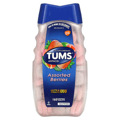

SCIENTIFIC SCORE
No researches found
N/A
USERS' SCORE
Excellent
Based on 1 Reviews
9.5
American Health Ester-C with Citrus Bioflavonoids
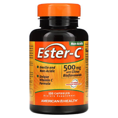
SCIENTIFIC SCORE
No researches found
N/A
USERS' SCORE
Excellent
Based on 1 Reviews
9.5
NOW Foods B-100
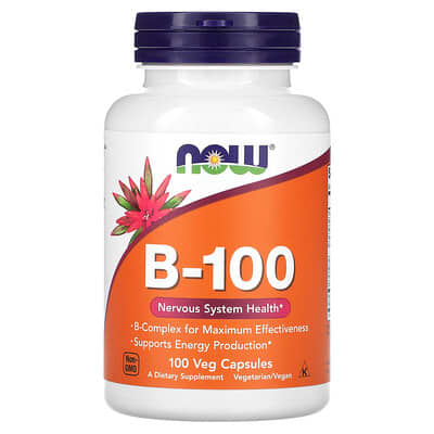
SCIENTIFIC SCORE
No researches found
N/A
USERS' SCORE
Excellent
Based on 1 Reviews
9.5
Medical Researches
SCIENTIFIC SCORE
Most Likely Effective
Based on 31 Researches
7.7
9
Eicosapentaenoic acid aids liver health
Antarctic Krill Oil Supplementation Attenuates Hypercholesterolemia, Fatty Liver, and Oxidative Stress in Diet-Induced Obese Mice.
Moderate relevance to liver disease
We explored the effectiveness of eicosapentaenoic acid (EPA), a key component of Antarctic krill oil, in battling obesity and its associated liver issues. Our investigation specifically aimed to understand how EPA influences cholesterol levels and overall liver health, especially in the context of diet-induced obesity.
Using a mouse model and analyzing various molecular pathways, we observed that a high-fat diet led to increased oxidative stress and obesity-related indicators, which are harmful to liver function. However, the introduction of EPA showed promising results in reducing oxidative stress, fat accumulation, and improving key metabolic parameters. These improvements were linked to better cholesterol management and support for liver health.
The findings suggest that EPA might serve as a valuable intervention for those struggling with obesity-related liver disease. By enhancing cholesterol metabolism and addressing oxidative stress, EPA could play a role in the prevention and treatment of these conditions. Overall, our results indicate a potential pathway for therapeutic applications in liver health through EPA supplementation.
Read More
9
Tyrosol positively impacts liver health
Tyrosol regulates hepatic lipid metabolism in high-fat diet-induced NAFLD mice.
Relevant findings on liver disease
We conducted an intriguing study to explore the effects of tyrosol (TYR), a compound enriched with beneficial properties, on nonalcoholic fatty liver disease (NAFLD) in mice. Males of the C57BL/6J strain were divided into groups that received either a low-fat diet, a high-fat diet, or a high-fat diet supplemented with 0.025% TYR for 16 weeks.
Observations revealed that the mice consuming the TYR-enriched diet experienced a notable decrease in both final body weight and liver fat accumulation compared to those on just the high-fat diet. A closer examination of liver metabolites showed an increase in key substances, including eicosapentaenoic acid (EPA), which suggests that TYR positively influences lipid metabolism and supports liver health.
We further investigated the mechanism behind these benefits and found that TYR interacts with a receptor known as peroxisome proliferator-activated receptor-alpha (PPARα). This interaction is crucial in regulating liver lipid processing, helping to turn on the genes that promote better lipid management.
Overall, this compelling evidence indicates that TYR, particularly through its role involving EPA and PPARα, could be a promising dietary addition for alleviating fatty liver disease in contexts of poor diets. We are excited about these insights and their potential implications for improving liver health.
Read More
9
Eicosapentaenoic Acid Reduces Hepatic Inflammation
Prevention of colitis-induced liver oxidative stress and inflammation in a transgenic mouse model with increased omega-3 polyunsaturated fatty acids.
Study suggests EPA's potential benefits
We explored the impact of eicosapentaenoic acid (EPA) and other omega-3 polyunsaturated fatty acids (n-3 PUFA) on liver inflammation related to inflammatory bowel disease (IBD). Using a transgenic mouse model known as fat-1 mice, we saw how increased levels of n-3 PUFA in tissues could play a role in reducing liver damage associated with colitis.
Our study pointed out that those fat-1 mice experienced less severe liver inflammation and oxidative stress compared to wild-type mice when subjected to a chemical that induces colitis. This is significant because while many discussions around omega-3 fatty acids center on their benefits for gut health, our findings suggest they also hold promise in addressing liver complications that might arise due to gut inflammation.
Additionally, we observed notable increases in certain beneficial metabolites derived from EPA, which are linked to reducing inflammation. These findings underline a strong connection between dietary n-3 PUFA intake and less oxidative stress in the liver, which could open up new avenues for therapeutic approaches in managing IBD and its systemic effects.
Read More
9
DHA combined with MCTs beneficial
Cosupplementation with DHA and medium-chain triglycerides ameliorates NAFLD and reduces amyloid-β accumulation by modulating hepatic lipid metabolism in APP/PS1 mice.
Moderately relevant due to combination
We explored the effects of docosahexaenoic acid (DHA) on liver disease, specifically looking at its role in nonalcoholic fatty liver disease (NAFLD) and its potential connection to Alzheimer's disease. Our study conducted on APP/PS1 mice involved four groups of animals fed different diets—one with DHA, one with medium-chain triglycerides (MCTs), and one that combined both treatments.
Throughout the study, which lasted eight months, we observed a significant reduction in blood and liver lipids in the group that received both DHA and MCTs. This combination not only alleviated signs of NAFLD but also reduced the buildup of amyloid-β (Aβ), a protein linked to Alzheimer's, in the brain and serum.
Additionally, our findings indicated that DHA combined with MCTs improved the activity of liver enzymes critical for lipid metabolism. This suggests that these compounds together may enhance the liver's ability to clear fat and cholesterol while also increasing Aβ clearance.
While we noted the benefits of DHA, it's important to remember that the effects we observed were influenced by the combination with MCTs, making it challenging to pinpoint DHA's isolated impact. Still, our research provides valuable insight into how enhancing dietary fats could support liver health and potentially mitigate connections to neurodegenerative diseases.
Read More
9
Docosahexaenoic acid for liver cancer
APT imaging of hepatocellular carcinoma signals an effective therapeutic response in advance of tumor shrinkage.
Examines DHA effectiveness in therapy
We explored the effectiveness of docosahexaenoic acid (DHA), particularly when delivered via nanoparticles, in treating liver disease, specifically hepatocellular carcinoma (HCC), in three rodent models. Our focus was on understanding how DHA affects HCC lesions as well as the performance of weighted amide proton transfer (APT) MRI as a monitoring tool.
In all three models—diethylnitrosamine (DEN) induced HCC, N1S1 syngeneic orthotopic xenograft, and human HepG2 ectopic xenograft—the APT MRI revealed higher signals from the cancerous tissue compared to surrounding normal tissue. Notably, in the DEN model, we found that the APT signal could effectively differentiate between malignant lesions and benign nodules.
After administering LDL-DHA nanoparticles directly into tumors, we observed a rapid decrease in APT signals within 72 hours, suggesting a promising therapeutic response. This trend was consistent in both N1S1 and HepG2 xenografts, indicating that DHA's effects, accelerated by nanoparticles, hold potential for therapeutic applications in liver cancer management.
Overall, our findings underscore the utility of APT imaging in the diagnostic and therapeutic landscapes of HCC, showcasing how innovative delivery methods can enhance treatment outcomes.
Read More
User Reviews
USERS' SCORE
Very Good
Based on 1 Reviews
8.5
8.5
Reduced inflammation
Perfectly Omega 3 aids in lowering the risk of chronic diseases such as heart disease, arteriosclerosis, and liver disease by diminishing inflammation in the body. The capsules are of average size, and the quality is excellent.
